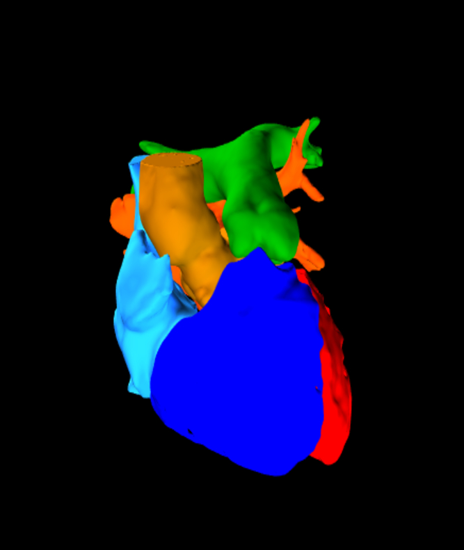
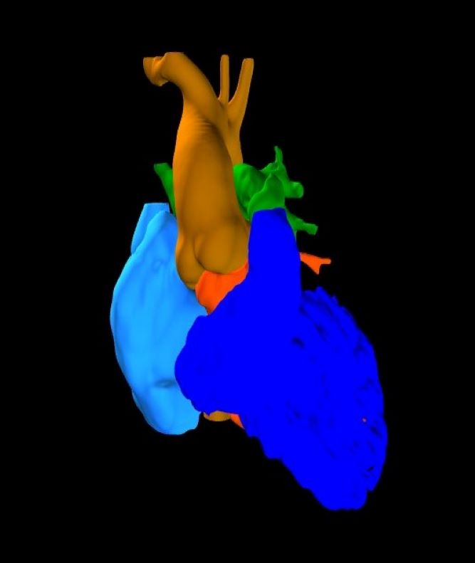
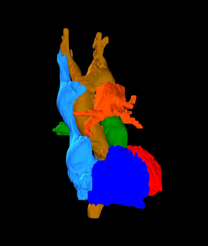
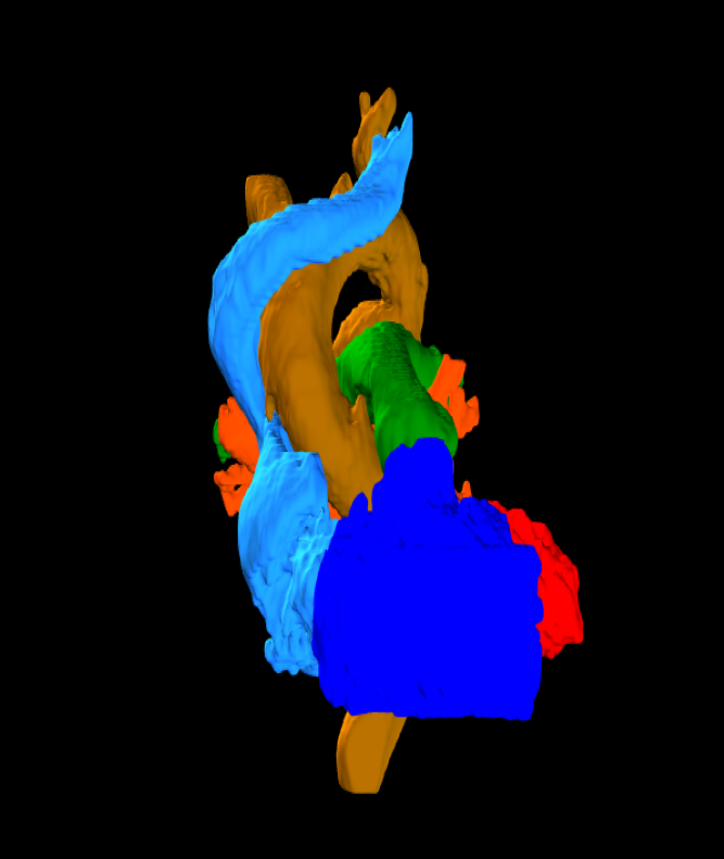
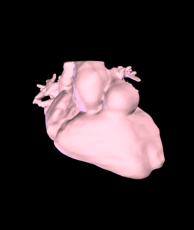
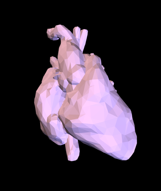
A growing collection of educational models created from cardiac-gated CT data of adult hearts, including from individuals with congenital heart disease. Segmentation: itk-SNAP and 3DSlicer. Textures: Blender.
This project is a collaboration by the Department of Anesthesia and Pain Management, Joint Department of Medical Imaging, Adult Congenital Cardiac Clinic, Electrophysiology, and The Division of Cardiac Surgery at Toronto General Hospital. Financial support from the UHN Foundation, and a Continuing Professional Development Award from the Faculty of Medicine at the University of Toronto.
Developed by: Azad Mashari MD, Joshua Qua Hiansen MSc, Kate Kazlovich, Ryan Ramos MD, Sachin Khargie, Mini Pakkal MD, Eitan Aziza MD, Massimiliano Meineri MD
A special thanks to Drs. M. Osten, E. Horlick, E. Hicky, K. Nair and RJ Cusimano for their support of the project.
 This work is licensed under a Creative Commons Attribution 4.0 International License. Model files (printable) can be found here. 3D prints can also be purchased here.
Toronto Heart Atlas by APIL on Sketchfab
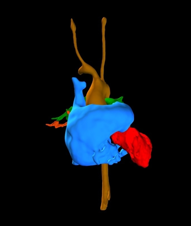
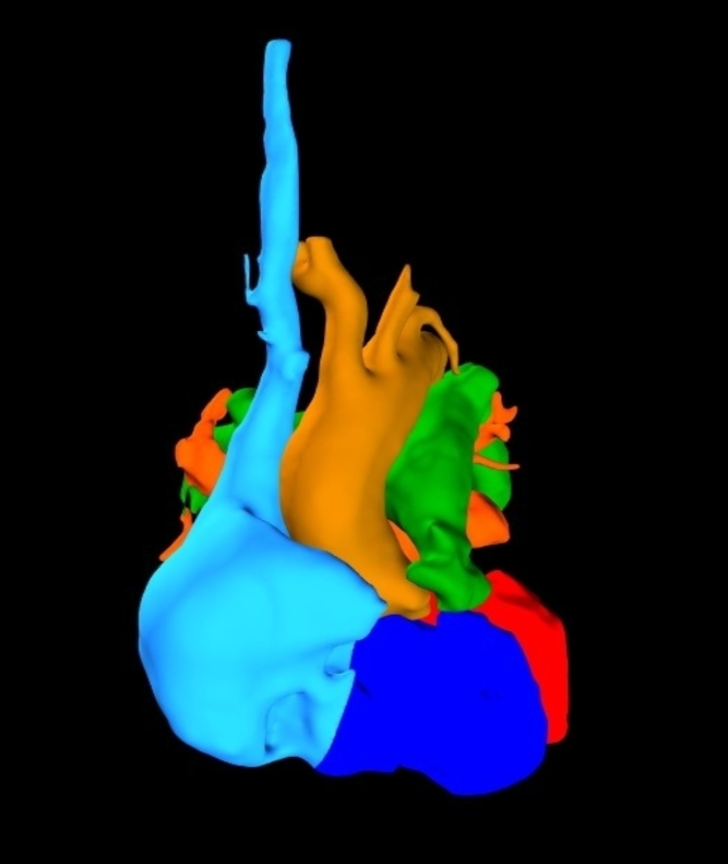
Diagnosis
- Hypoplastic right heart: tricuspid atresia, pulmonary stenosis, hypoplastic right ventricle
- Multiple VSDs
- Secundum ASD
Procedures
- Right-sided BTT shunt
- Bjork Fontan operation; ligation of BT shunt
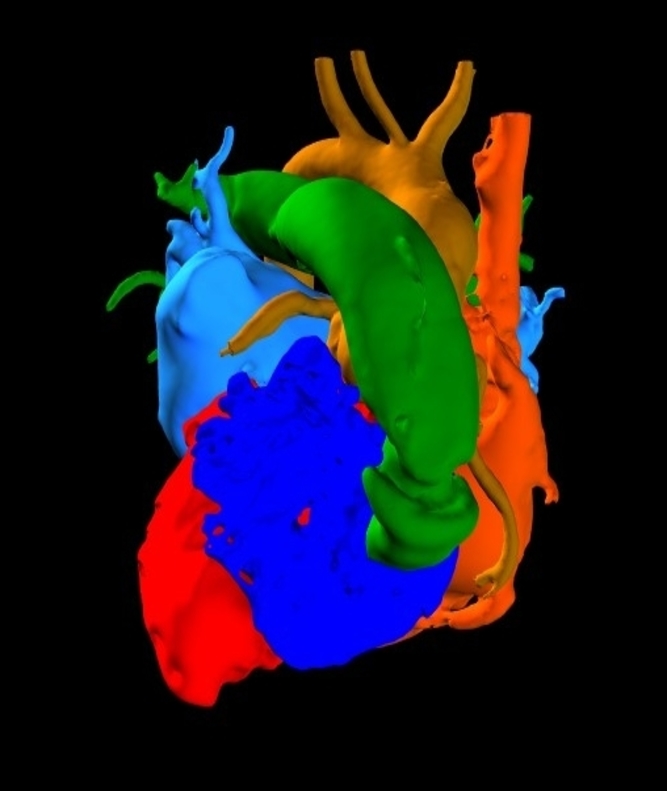
Diagnosis
- Tetralogy of Fallot, (uncorrected): Overriding aorta, pulmonary stenosis, subaortic ventricular septal defect, right ventricular hypertrophy
Procedures
- Remnant of BTT shunt can be seen from left subclavian artery to left pulmonary artery.
- No further correction until age 65. Underwent pulmonary valve replacement subsequently.
Diagnosis
- Orientation: Situs inversus totalis; dextrocardia; Infero-posterior morphologic LV, supero-anterior morphologic RV; Right aortic arch with mirror-image branching
- Segmental anatomy: Complex TGA: Concordant A-V, discordant V-A connections: posterior pulmonary valve, anterior aortic valve, with ventricular L-loop
- Shunts: Secondum ASD; Membranous VSD
- Pulmonary stenosis; subpulmonary outflow tract obstruction.
Procedures
- Patch closure of membranous VSD, tunnel-like connection of LV to aorta; pulmonary valvotomy and excision of fibrous tissue from the RVOT, closure of secundum ASD;
- Removal of VSD patch, Morrow-like incision on the ventricular septum; closure of VSD, Relief of subpulmonic outflow obstruction; transvenous atrial and ventricular pacemaker insertion
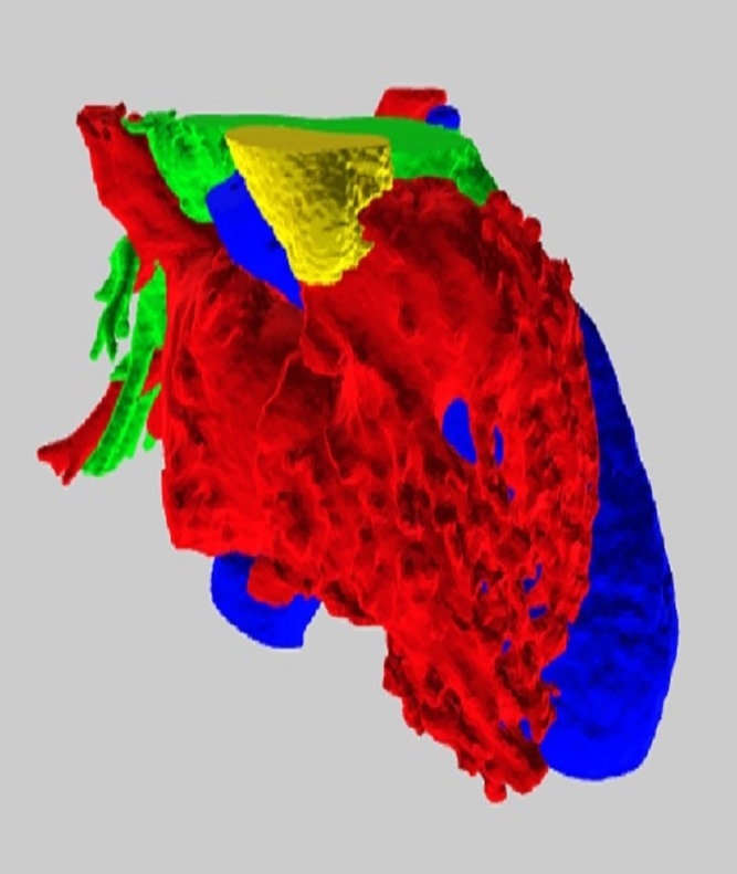
Diagnosis & Procedures
- D-TGA (AV concordance; VA discordance) post atrial switch (Mustard) repair.
- Large leak between IVC and pulmonary venous baffle.
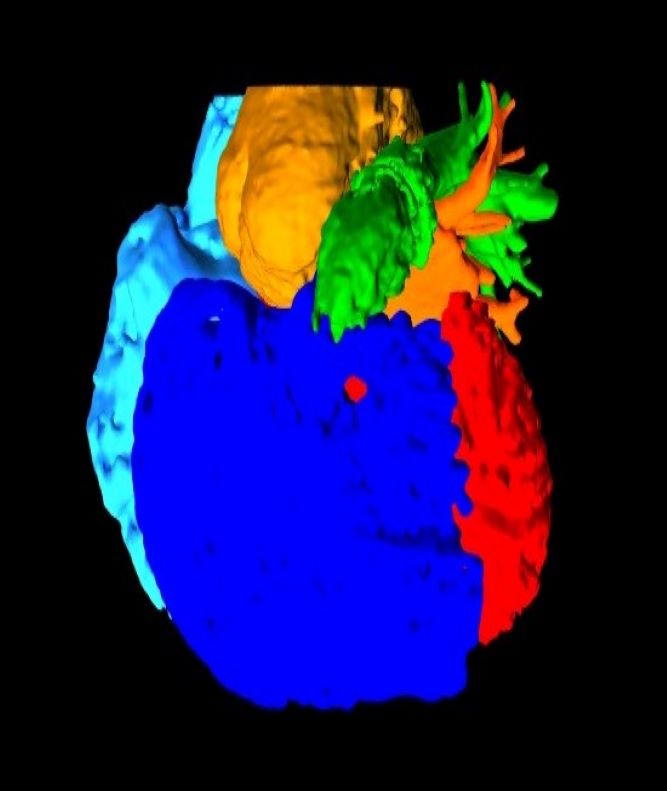
Diagnosis & Procedure
- Hypoplastic left heart; aortic atresia
- Ventricular septal defect.
- Yasui repair
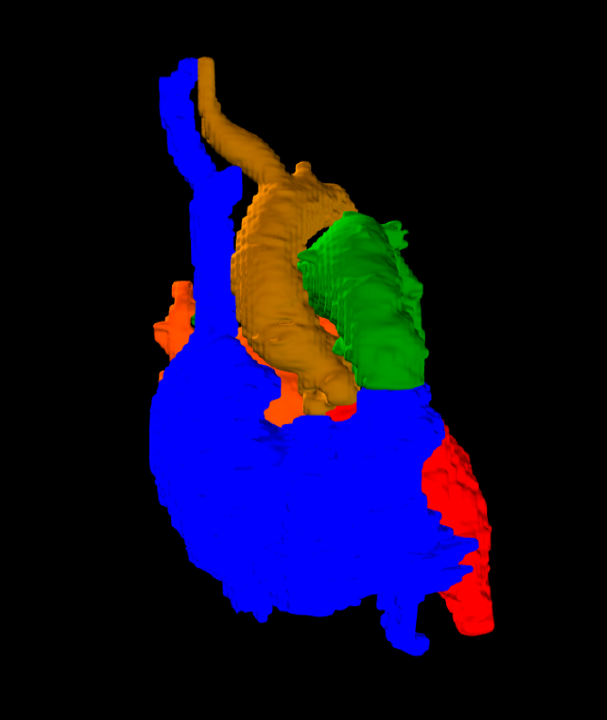
Heart: 2018014-01
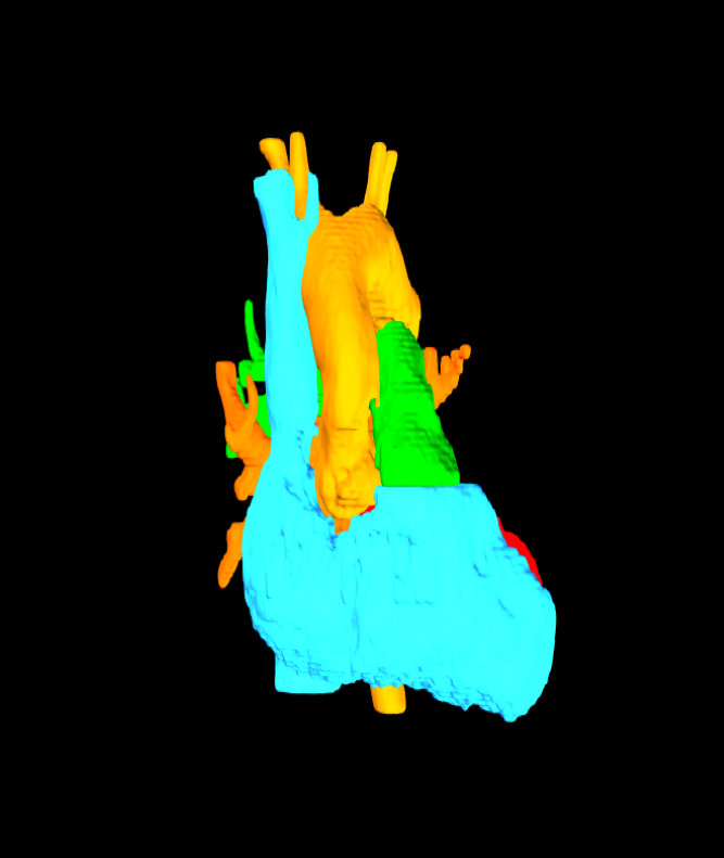
Heart: 2018015-01
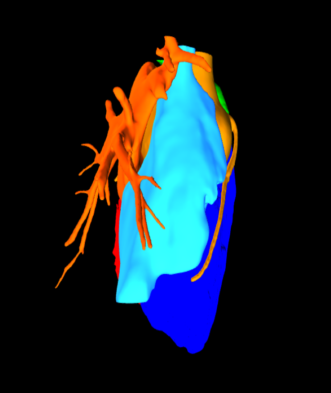
Hearts: 2018016-01 Marfan syndrome aortic aneurysm
Aortic root aneurysm in patient with Marfan syndrom
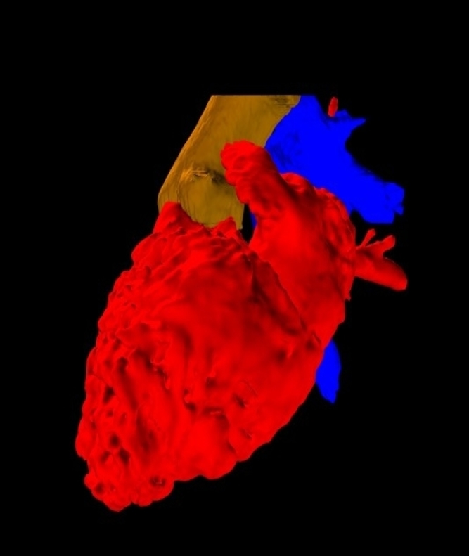
Heart: 2018017 d-TGA Hypoplast RV Lat Tunnel Fontan
Male Age 25-30. Source Cardiac CT. Software: ITK-SNAP, 3D Slicer, Blender, Meshmixer.
Original Diagnosis
- Situs solitus, D-TGA: A-V concordance; V-A discordance
- Double inlet left ventricle; hypoplastic right ventricle
- Pulmonary stenosis
- Subvalvular aortic stenosis
- Atrial septal defect
Procedures
- Right BT shunt (First year)
- Bidirectional cavopulmonary anastomosis, aortic myomectomy (Age 6)
- Lateral tunnel Fontan (Age 6)
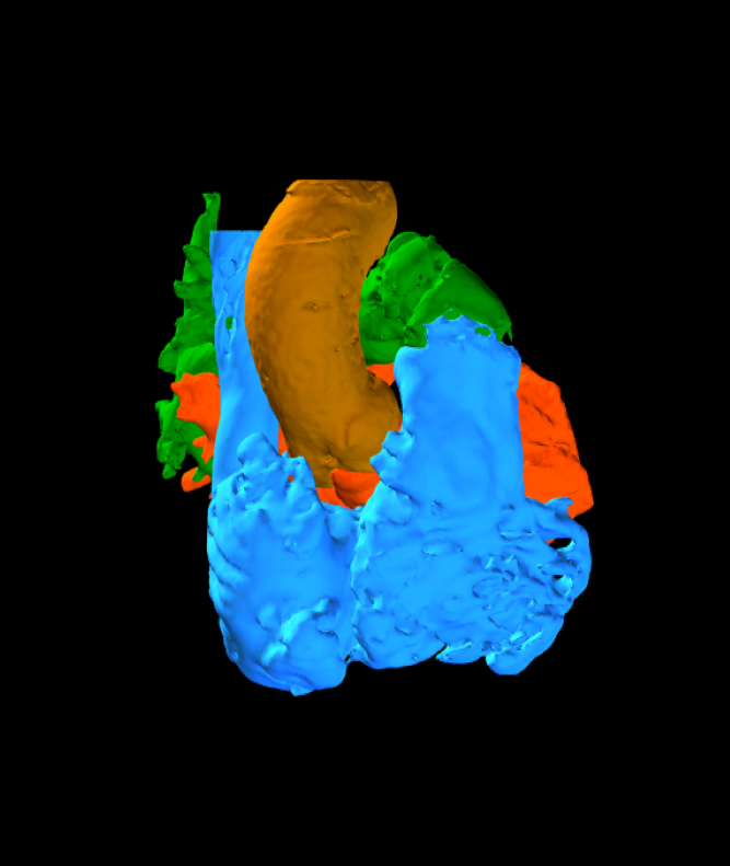
Heart: 2018018-01
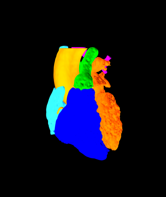
Heart: 2018019-01
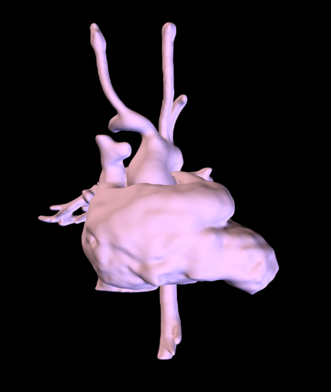
Myocardium: 2018001-01
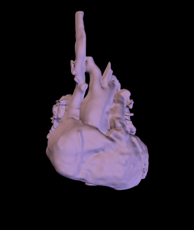
Myocardium: 2018002-01
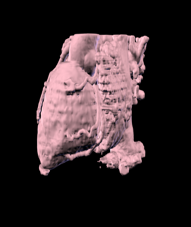
Myocardium: 2018003-01
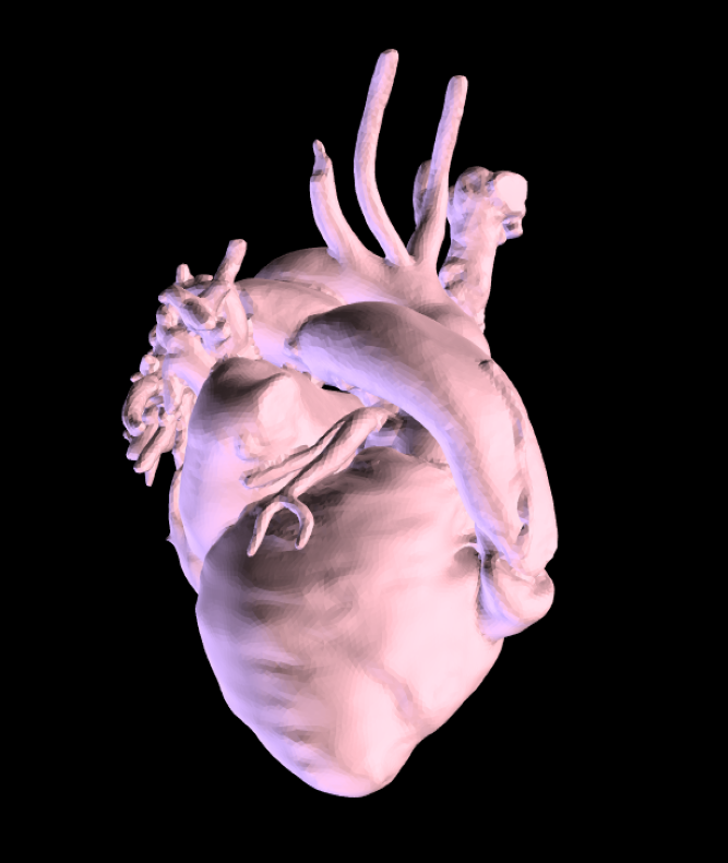
Myocardium: 2018003-02
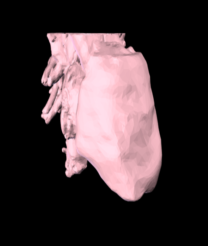
Myocardium: 2018004-01
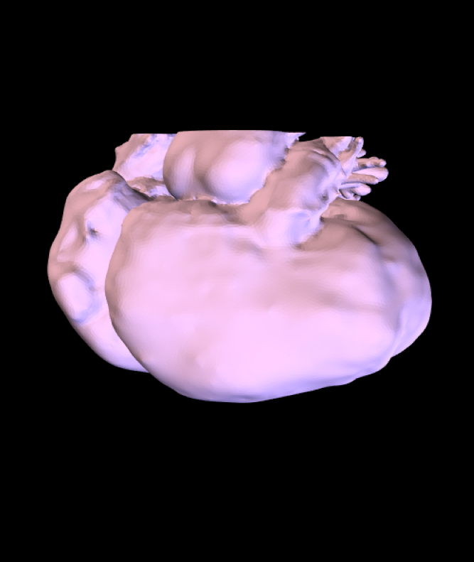
Myocardium: 2018005-01
