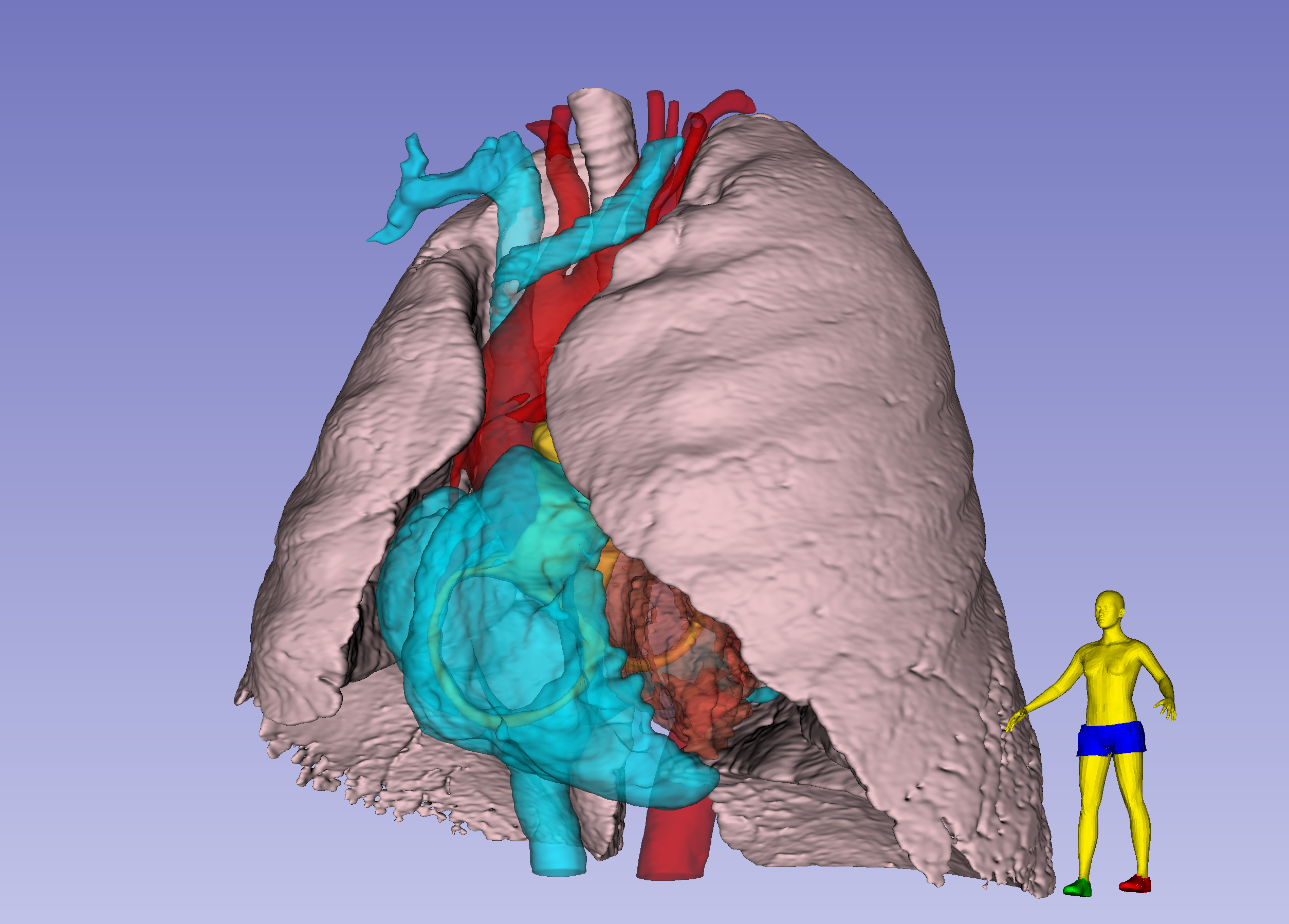
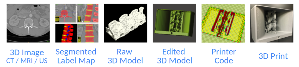
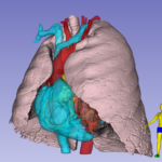
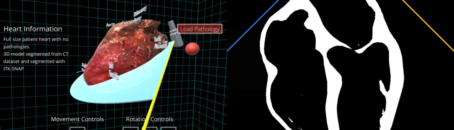
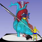
Interactive rendering of lung and heart blood pools
for teaching TTE
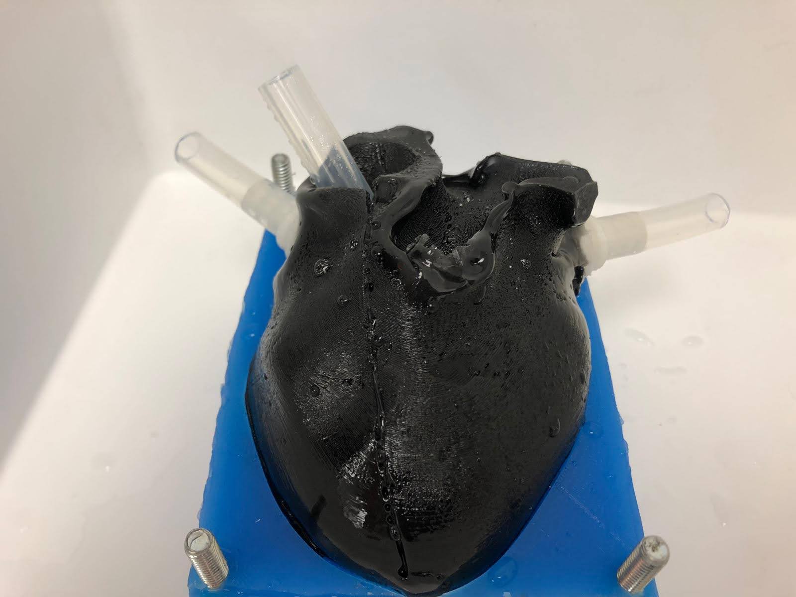
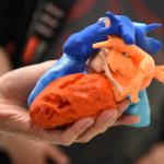
Cast Heart for
Echocardiography phantom
CT data. FDM printed positives, Silicone molds; Gel wax and ballistic gel casts with graphite.
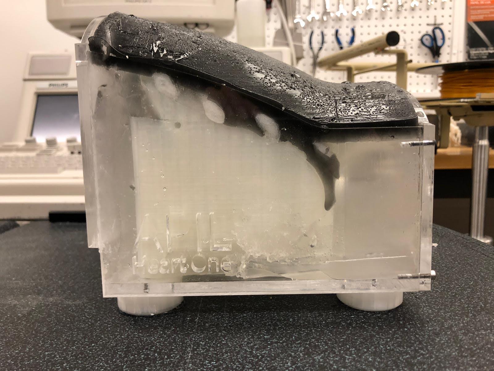
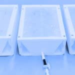
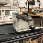
Echocardiography phantom
CT data. FDM printed positives, Silicone molds; Gel wax and ballistic gel casts with graphite. Lasercut case.
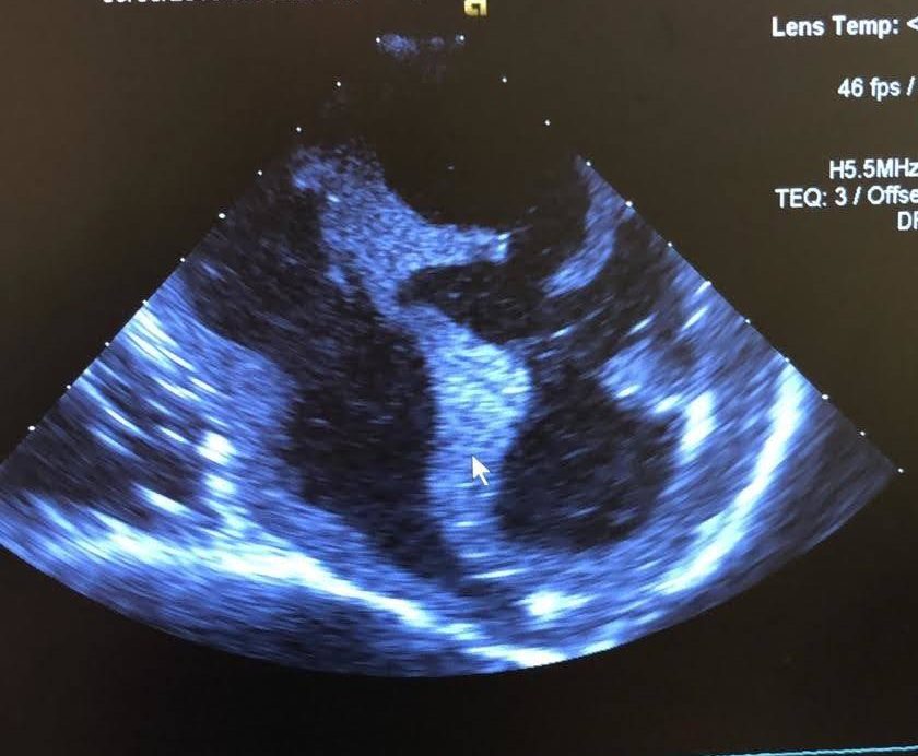
Ultrasound Image from
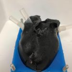
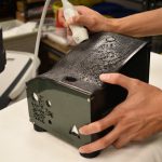
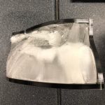
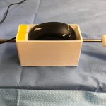
Echocardiography phantom
CT data. FDM printed positives, Silicone molds; Gel wax and ballistic gel casts with graphite. Lasercut case.
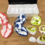

Echocardiography phantom
CT data. FDM printed positives, Silicone molds; Gel wax and ballistic gel casts with graphite. Lasercut case.
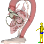
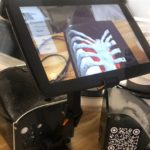
Augmented Reality (AR)
projection of heart and ribcage on echocardiography phantom.
Cardiac CT.

Interactive rendering
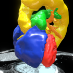
Toronto Heart Atlas
Normal Morphology; Type-A Aortic Dissection
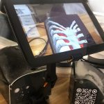
Ultrasound-guided subclavian central venous access task trainer
CT data. FDM printing. Silicone and gel casting. Lasercut case.
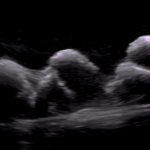
Ultrasound Image from
Echocardiography phantom
CT data. FDM printed positives, Silicone molds; Gel wax and ballistic gel casts with graphite. Lasercut case.
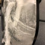
Echocardiography phantom
CT data. FDM printed positives, Silicone molds; Gel wax and ballistic gel casts with graphite. Lasercut case.

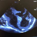
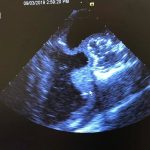
Ultrasound Image from
Echocardiography phantom
CT data. FDM printed positives, Silicone molds; Gel wax and ballistic gel casts with graphite. Lasercut case.

Toronto Heart Atlas
Complex TGA; Situs inversus; Dextrocardia. Post insertion of RV to PA conduit.
Cardiac CT. FDM Print.
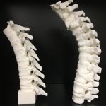
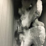
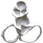
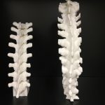
Spine models
for development of neuraxial anesthesia phantom
CT. FDM Print.
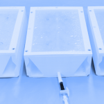
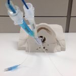
Ultrasound-guided vascular access phantom
CAD. FDM printing. Gel casting. Lasercut case.
Ultrasound-guided subclavian central venous access task trainer
CT data. FDM printing. Silicone and gel casting. Lasercut case.
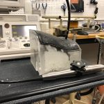
Lung-isolation Training Phantom
CT. FDM print.
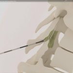
Thoracic epidural insertion
CT. Educational Animation.
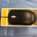
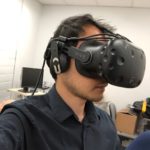
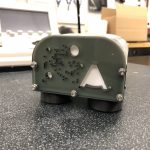
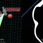
TEE simulator controller
Using 2-button wheel-mouse simulates change of probe depth, omniplane angle and probe flexion.
Spine models
for development of neuraxial anesthesia phantom
CT. FDM Print.
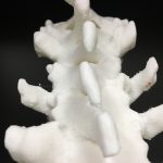
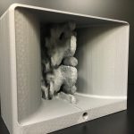
Spine model - Scoliosis
for development of neuraxial anesthesia phantom
CT. FDM Print.

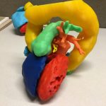
FOCUS Heart Models
showing standard imaging planes for FOCUS TTE
Cardiac CT. FDM Print. Magnets

TEE simulator controller
Using 2-button wheel-mouse simulates change of probe depth, omniplane angle and probe flexion.